
Biology Today and Tomorrow without Physiology (MindTap Course List)
5th Edition
ISBN: 9781305117396
Author: Cecie Starr, Christine Evers, Lisa Starr
Publisher: Cengage Learning
expand_more
expand_more
format_list_bulleted
Concept explainers
Videos
Textbook Question
Chapter 3, Problem 1VQ
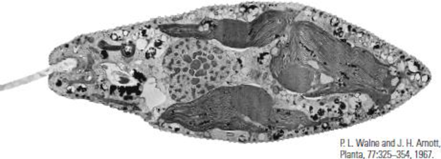
What type of micrograph is shown below? Is the organism pictured prokaryotic or eukaryotic? How can you tell?
Expert Solution & Answer
Want to see the full answer?
Check out a sample textbook solution
Students have asked these similar questions
Carefully observe the cell morphology of the two organisms below. Then, draw the cell morphology. Cells should be drawn large enough so that you can show an accurate shape and arrangement. Your drawing should capture the relative length and width of the cells. There should be no pointy corners, no open circles, no extra “tails” and no overlapping rings. Specify cell morphology and if the organism a) Escherichia coli or b) Staphylococcus aureus
Note :
— don't copy in google or bartleby. Plagarize checker will be used.
QUESTION :
Differentiate the Cell Membrane of Gram Positive Bacteria and Gram Negative Bacteria.
Measure the length of Cell X using the ruler in Microsoft Word. Assuming the actual length to be 3 um, calculate the magnification. Show your complete solution.
Based on Figure 1, what internal organization can be distinguished in cell X? Why?
Based in Figure 1, can you see a limiting membrane? Can you deduce its presence? Why or why not?
Figure 2 is an electron micrograph of the same type of bacterium as shown in Figure 1. The picture has been obtained by cutting a very thin section of the bacterial cell along its longitudinal axis.
Measure the total length of the cell, and assuming the actual length to be 2.1 um, calculate the magnification.
With reference with Figure 2, what are the major differences between the inclusions found in Figure 1 and the way they appear in Figure 2?
What other structural features can be resolved?
Chapter 3 Solutions
Biology Today and Tomorrow without Physiology (MindTap Course List)
Ch. 3 - Prob. 1FIOCh. 3 - Organelles and Cystic Fibrosis A plasma membrane...Ch. 3 - Organelles and Cystic Fibrosis A plasma membrane...Ch. 3 - Prob. 3DIDCh. 3 - All cells have these three things in common: a....Ch. 3 - Unlike eukaryotic cells, prokaryotic cells...Ch. 3 - Every cell is descended from another cell. This...Ch. 3 - Prob. 4SQCh. 3 - True or false? Some protists start out life with...Ch. 3 - Cell membranes consist mainly of ________ and...
Ch. 3 - Prob. 7SQCh. 3 - In a lipid bilayer, the of all the lipid molecules...Ch. 3 - The main function of the endomembrane system is...Ch. 3 - Enzymes contained in __________ break down...Ch. 3 - Put the following structures in order according to...Ch. 3 - No animal cell has a ______. a. plasma membrane b....Ch. 3 - Prob. 13SQCh. 3 - Prob. 14SQCh. 3 - Match each cell component with its main function.Ch. 3 - In a classic episode of Star Trek, a gigantic...Ch. 3 - Prob. 2CTCh. 3 - What type of micrograph is shown below? Is the...
Knowledge Booster


Learn more about
Need a deep-dive on the concept behind this application? Look no further. Learn more about this topic, biology and related others by exploring similar questions and additional content below.Similar questions
- Label the structures of the prokaryotic cell. Not all terms will be used.arrow_forwardWhat are bacteria? what is bacterial culture? how to grow bacteria and why? why are there so many bacteria on the keyboard?arrow_forwardUsing bright-field microscopy to look at a slide prepared with a basic dye you observe cells with a clear inner compartment within the cell at 400X magnification. The cell is most likely a(n) A) prokaryote. B) bacterium. C) archaeon. D) eukaryote.arrow_forward
- The Gram staining technique is useful to classify Bacteria. Can the technique be applied to Archaea? Discuss briefly.arrow_forwardWhat is not part of a Bacterial cell base on the picture?arrow_forwardWhich of the following microscopes typically requires the use of vital dyes (like methylene blue) to visualize large subcellular structures in a living cell (like the nucleolus or the mitochondrion)? the scanning/tunneling electron microscope (STEM), with good resolution up to about 100,000,000x the transmission electron microscope (TEM), with good resolution up to about 100,000x the scanning electron microscope (SEM), with good resolution up to about 1,000,000x the compound light microscope, with good resolution up to about 1,500x all of the above microscopes would be equally useful in visualizing the interior of organellesarrow_forward
- What are 5 cellular parts that are specific to ALL bacteria. This should not include any cellular parts listed above (like cytoplasm) that both prokaryotes and eukaryotes will have. This also should not include cellular parts that only specific types of bacteria will have (example: flagella). Then state the function of the cellular part (what does it do for the cell?)arrow_forwardThe following chart lists a variety of cell types and classifies them by their cell arrangement and Domain. Which number is incorrect? Number Cell Type Cell Arrangement Domain 1 Prokaryotic Unicellular Bacteria Prokaryotic Multicellular Archaea 3 Eukaryotic Unicellular Eukarya 4 Eukaryotic Multicellular Eukarya O 1 O 2 0 3 4arrow_forwardwhat are three cellular parts that are specific to ALL bacteria? This should not include any cellular parts listed that both prokaryotes and eukaryotes will have. This also should not include cellular parts that only specific types of bacteria will have (example: flagella). Then state the function of the cellular part (what does it do for the cell?arrow_forward
- Rank the following in order from smallest to largest: ant, prokaryoticcell, actin molecule, microtubule, nitrogen atom. What type ofmicroscope (if any) would you need if you wanted to see each?arrow_forwardWhat is the process to perform a Gram Stain? What happens if you make a mistake on a step (Think about different mistakes you could make)? What are the proper results? What structures do Archaeans use to attach to surfaces (Look like treble hooks)? What are bacterial inclusions? What are the similarities and differences in Gram - cells and Gram + cell structure? What is the function of ribosomes and what are they mad up of? What component of bacterial cells helps to combat/regulate osmotic forces? What are Koch’s postulates? What are they used for? Explain the process of endospore formation in endospore-producing organisms. What are the major components of the bacterial & eukaryotic cell? What is/are a pilus/pili and what do microbes use them for? What is the prokaryotic flagellum made up of?arrow_forwardIn the diagram below, identify the structures of a cyanobacterial cell based on the following descriptions: a) Outer cellular covering which includes: Mucilaginous layer – outermost layer covering the cell wall; protects the cell from harmful factors of the environment Cell wall – found just below the mucilaginous layer; 2 or 3-layered, the inner layer lies in between the outer wall layer and plasma membrane; the outer layer is made of peptidoglycan Innermost plasma membrane – selectively permeable membrane enclosing the cytoplasm b) Cytoplasm – found below the plasma membrane; the protoplasm which contains structures of different shapes and functions. Lamellae, which contain pigments such as chlorophylls, carotenes, xanthophylls, phycoerythrin and phycocyanin, are located in the peripheral region of cytoplasm. Ribosomes may also be found scattered in the cytoplasm. c) Nucleic material – the nucleoplasm that is centrally located in the cell and contains chromatin in the form…arrow_forward
arrow_back_ios
SEE MORE QUESTIONS
arrow_forward_ios
Recommended textbooks for you
 Biology Today and Tomorrow without Physiology (Mi...BiologyISBN:9781305117396Author:Cecie Starr, Christine Evers, Lisa StarrPublisher:Cengage Learning
Biology Today and Tomorrow without Physiology (Mi...
Biology
ISBN:9781305117396
Author:Cecie Starr, Christine Evers, Lisa Starr
Publisher:Cengage Learning
Biology - Intro to Cell Structure - Quick Review!; Author: The Organic Chemistry Tutor;https://www.youtube.com/watch?v=vwAJ8ByQH2U;License: Standard youtube license