
Laboratory Manual For Human Anatomy & Physiology
4th Edition
ISBN: 9781260159363
Author: Martin, Terry R., Prentice-craver, Cynthia
Publisher: McGraw-Hill Publishing Co.
expand_more
expand_more
format_list_bulleted
Videos
Textbook Question
Chapter 2, Problem F2.11A
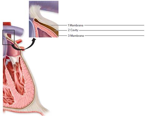
FIGURE 2.11 Label the specific serous membranes and cavities (1—3) of the heart.
Expert Solution & Answer
Want to see the full answer?
Check out a sample textbook solution
Students have asked these similar questions
NOTES
and the left side serving the systemic circuit. In Figure 16-15, complete
the schematic showing the blood flow to and from the heart (the starting
points are given to you). Use a blue pen or pencil to denote the direction of
deoxygenated blood and a red pen or pencil for oxygenated blood flow.
Include the names of the major vessels, chambers, and valves involved,
based on the following list:
lung capillary beds
body capillary beds
right ventricle
left ventricle
bicuspid valve
superior vena cava
tricuspid valve
inferior vena cava
pulmonary semilunar valve
pulmonary trunk
aortic semilunar valve
R. and L. pulmonary arteries
R. and L. pulmonary veins
aorta
Pulmonary Circulation
Systemic Circulation
Right atrium
Left atrium
URA--YCK
HAT
Lungs
Body
Figure 16-15. Schematic of circulation
Label the three views of the heart in Figure 12.1 with the following terms.
O Right atrium
O Left atrium
Great Vessels
O Superior vena cava
O Inferior vena cava
Structures of the Ventricles
O Right ventricle
O Left ventricle
O Interventricular septum
O Chordae tendineae
O Papillary muscles
O Pulmonary trunk
O Pulmonary veins
O Aorta
Coronary Vessels
O Right coronary artery
O Anterior interventricular
artery (left anterior
descending)
O Coronary sinus
O Great cardiac vein
O Circumflex artery
Atrioventricular Valves
O Tricuspid valve
O Mitral valve
Semilunar Valves
O Pulmonary valve
O Aortic valve
B
FIGURE 12.1 Heart: (A) anterior view;
(B) frontal section; (C) posterior view
Label the three views of the heart in Figure 12.1 with the following
O Right atrium
O Left atrium
Great Vessels
O Superior vena cava
O Inferior vena cava
Structures of the Ventricles
O Right ventricle
O Left ventricle
O Interventricular septum
O Chordae tendineae
O Papillary muscles
O Pulmonary trunk
O Pulmonary veins
O Aorta
Coronary Vessels
O Right coronary artery
O Anterior interventricular
artery (left anterior
descending)
O Coronary sinus
O Great cardiac vein
O Circumflex artery
Atrioventricular Valves
O Tricuspid valve
O Mitral valve
Semilunar Valves
O Pulmonary valve
O Aortic valve
C
Chapter 2 Solutions
Laboratory Manual For Human Anatomy & Physiology
Ch. 2 - The basis for communication in anatomy and...Ch. 2 - Which of the following is not a body cavity?...Ch. 2 - The pericardium is associated with the lung....Ch. 2 - The ____________ plane divides the body into left...Ch. 2 - The abdominopelvic cavity can be subdivided into...Ch. 2 - The larynx is part of the __________ system....Ch. 2 - The epigastric region is a portion of the _______...Ch. 2 - In the posterior view, the cubital region is...Ch. 2 - The brachial surface region pertains to the wrist....Ch. 2 - A frontal plane divides the body into anterior and...
Ch. 2 - Match the body cavities in column A with the...Ch. 2 - FIGURE 2.10 Label body cavities 1-5. Add a...Ch. 2 - FIGURE 2.11 Label the specific serous membranes...Ch. 2 - the organ systems in column A with the principal...Ch. 2 - Indicate whether each of the following sentences...Ch. 2 - Indicate whether each of the following sentences...Ch. 2 - Indicate whether each of the following sentences...Ch. 2 - Indicate whether each of the following sentences...Ch. 2 - Indicate whether each of the following sentences...Ch. 2 - Indicate whether each of the following sentences...Ch. 2 - Indicate whether each of the following sentences...Ch. 2 - Indicate whether each of the following sentences...Ch. 2 - Indicate whether each of the following sentences...Ch. 2 - Indicate whether each of the following sentences...Ch. 2 - Indicate whether each of the following sentences...Ch. 2 - Indicate whether each of the following sentences...Ch. 2 - FIGURE 2.12 Label the planes of the sectioned...Ch. 2 - Figure 2.13 Label the indicated body surface...Ch. 2 - Examine the letters A, B, and C indicated on...
Knowledge Booster


Learn more about
Need a deep-dive on the concept behind this application? Look no further. Learn more about this topic, biology and related others by exploring similar questions and additional content below.Similar questions
- In this view of the superior surface of the heart, at the base where all the blood vessels enter and leave the heart, #17 is the opening where the______ enter and transport blood into the _______. (specific chamber)arrow_forwardThe heart is lateral to the lungs. True or falsearrow_forwardCould you draw a picture of the flow of blood through the heart. And label at least 10 chambers,vessels, or structures. Could you also in the drawing include colors indicate which chambers or vessels are carrying oxygenated (red) blood or deoxygenated blood (blue)arrow_forward
- HQ.2 Label the chambers and valves seen in an anterior view of the heart.arrow_forwardDraw the posterior view of the heart exposing the coronary vesselsarrow_forwardtrace a drop of blood from the left ventricle of the heart to the wrist of the right hand and back to the heart(name each vessel) then trace the drop of blood to the dorsum of the right foot and back to the right side of the heart.arrow_forward
- On a diagram of a frontally sectioned heart, indicate the location of the cardiac skeleton.arrow_forwardIf a drop of blood were going from the inferior vena cava to the heart, trace the path of that drop of blood from the vessel (IVC) through the four chambers of the heart, four valves of the heart and the lungs, in the correct sequence, ending with the drop of blood going into the aorta.arrow_forwardLabel the structures to the letter A- ____________ sac B- ___________ vena cava C- ___________ trunk D- ___________ arteries E- ___________ veins F- ___________ arch= G- thoracic ________ H- abdominal _______ I- J-arrow_forward
- A pulse can be felt in the following arteries: superficial temporal, facial, common carotid, brachial, femoral, popliteal, posterior tibial, and dorsalis pedis (Figure 20.8b). Name the artery from which each artery listed branches. Here is an example to get you started: The superficial temporal artery is a branch of the external carotid artery.arrow_forwardIn the image attached help me locate the anterior ventricular artery and draw a thrombosis in that artery and then shade the area of the heart that would experience a myocardial infarction. Thanksarrow_forwardfill in the blanks Blood that has circulated to the heart muscle itself also returns to the right atrium. Once blood leaves the cells of the myocardium, it flows into a system of cardiac veins, eventually flowing into the great cardiac vein, and then the coronary sinus. On this anterior view of the heart, locate the great cardiac vein and the coronary sinus (shown through to the back). Slide 14: Identify the two large veins that merge and flow into the superior vena cava: A._________________________________ and B._________________________________. (Note that unlike the arterial system, there are two veins, a right and a left, with this name.) Locate either the right or left brachiocephalic vein. Each is formed by the union of two slightly smaller veins. An C.___________________________ vein draining the head and a D.______________________ vein bringing blood back from the arm. Locate an additional vein draining blood from the head that flows into the subclavian vein, the…arrow_forward
arrow_back_ios
SEE MORE QUESTIONS
arrow_forward_ios
Recommended textbooks for you
 Human Physiology: From Cells to Systems (MindTap ...BiologyISBN:9781285866932Author:Lauralee SherwoodPublisher:Cengage Learning
 Biology 2eBiologyISBN:9781947172517Author:Matthew Douglas, Jung Choi, Mary Ann ClarkPublisher:OpenStax
Human Physiology: From Cells to Systems (MindTap ...
Biology
ISBN:9781285866932
Author:Lauralee Sherwood
Publisher:Cengage Learning
Biology 2e
Biology
ISBN:9781947172517
Author:Matthew Douglas, Jung Choi, Mary Ann Clark
Publisher:OpenStax
Respiratory System; Author: Amoeba Sisters;https://www.youtube.com/watch?v=v_j-LD2YEqg;License: Standard youtube license