
Concept explainers
Figure 18.13 Are you able to recognize and identify the various formed elements? You will need to do this is a systematic manner, scanning along the image. The standard method is to use a grid, but this is not possible with this
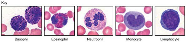
Figure 18.13 Leukocytes (Micrographs provided by the Regents of University of Michigan Medical School © 2012)

Trending nowThis is a popular solution!

Chapter 18 Solutions
Anatomy & Physiology
Additional Science Textbook Solutions
College Physics
Human Anatomy & Physiology (11th Edition)
Microbiology: An Introduction
Campbell Biology in Focus
Campbell Essential Biology with Physiology (5th Edition)
Microbiology: An Introduction (13th Edition)


- I will make your life a bit easier and not make you count four additional grids. So let's assume this was a random, relatively uniform sample and arbitrarily assign the other four squares to the exact same number of red blood cells. What is the Total red blood cell count? Calculate it as (#RBC you counted *5)*10,000 Is you answer normal? Edit View Insert Format Tools Table Paragraph v | в I U 12pt varrow_forwardIn hematology laboratory, automation has been the ongoing trend in terms of cell sorting and identification. Do you think it is about time to phase out the manual method of blood cell counting and identification in this area? Why or why not?arrow_forwardCreate a biological drawing of the blood films below (A and B). Label one example of each of the following: red blood cells, leukocytes, debris, staining artefacts, dead cells, empty space.arrow_forward
- Can all abnormal hemoglobins be diagnosed by electrophoresis? Explain your answer in detail.arrow_forwardYou are performing an experiment to diagnose three patients who are ill. In a hematology lab, you run a CBC using the hematology analyzer on all 3 of your patience blood samples, once you collect that data, you then conduct a blood smear/stain to get a better look at the 3 patients blood cells and compare those results to those of the CBC report. You conclude that patient 1 has malaria, patient 2 has sickle cell anemia, and patient 3 has hemophilia. Can you Identify your independent, dependent and controlled variables?arrow_forwardplease fill in the charts as the directions states i have included the 2 pictures you need to look at in order Arrange the key and blood smear side by side so that both are visible Use the tally charts below to record the counts for your patient as you go. To count: Start in the upper left corner of the first photo. Identify the white blood cell in the first square and put a mark by its corresponding name on the tally chart. Repeat this process as you move across to the next square and then the next square and so forth. If a square has two WBCs, record both types. There are two sheets of cells to count. Once you have counted all the squares you should have a total of 100 cells. Total the tally for each cell type. Add your total tally for each cell type together to be sure you have a total of 100 recorded. Suggestion: Do not go through and count all the neutrophils, then go through and count all the lymphocytes, then go through and count all the monocytes, etc. Go through the…arrow_forward
- In Figure 1.16, blood samples are taken from a gate (U), an injured woman (S1), and her former husband (S2). The woman claimed that she as attacked by her former husband. The man had a cut on his hand, which he explained as a cut from a broken water glass. Is the unknown sample from her wound, from her former husband, or from an unknown third person? Answer: The unknown sample is from her wound. -From the same case, a second blood sample was collected from a stain on the floor of the former husband's home (Figure 1.17). Is this stain from the woman, her former husband, or an unknown third person? Answer: The stain on the floor is from her former husband. A third blood sample was collected from a knife in the alley two blocks from the woman's home (Figure 1.18). Is this stain from the woman, her former husband, or an unknown third person? Answer: It is from an…arrow_forwardIn Medtrix Laboratory, Jane, a medical technologist, does a manual reticulocyte counting for Patient A using Miller Disc. Jane was able to count 25 Reticulocytes in the large square and 112 RBCs in the small square. The attached photo is Patient A’s Complete Blood Count result. Using this data, how many reticulocytes are there in 1L of the patient’s blood? Does it fall under the reference interval?arrow_forwardin these two images identify if there are platelets and nRBcarrow_forward
- Create a biological drawing of the blood film below. Label one example of each of the following: red blood cells, leukocytes, debris, staining artefacts, dead cells, empty spacearrow_forwardUse Figure 37.2 to identify the leukocytes in the micrograph on the left. __ neutrophil ___ lymphocyte __ eosinophil ____ basophilarrow_forwardIn one square of a hemacytometer you count 55 cells. What is the approximate concentration of cells per l (liter) in this sample? (note that "^" means superscript, e.g. 10^2 means 10 to the 2nd power) The options are 5.5 X 10^5 2.75 X 10^8 5.5 X 10^8 2.75 X 10^5arrow_forward
 Anatomy & PhysiologyBiologyISBN:9781938168130Author:Kelly A. Young, James A. Wise, Peter DeSaix, Dean H. Kruse, Brandon Poe, Eddie Johnson, Jody E. Johnson, Oksana Korol, J. Gordon Betts, Mark WomblePublisher:OpenStax College